Research Article 
 Creative Commons, CC-BY
Assembly Formation of Synthetic DNA (E. coli) Crown Cells with Salt “Reconstruction and Regeneration of Synthetic DNA Crown Cells”
*Corresponding author: Shoshi Inooka, Japan Association of Science Specialists, Japan
Received: April 18, 2022; Published: April 26, 2022
DOI: 10.34297/AJBSR.2022.16.002200
Abstract
DNA crown cells (artificial cells) have an outer membrane covered with DNA and can be synthesized readily using sphingosine (Sph)-DNA-adenosine mixtures (synthetic DNA crown cells) and can proliferate within egg whites (DNA crown cells). Numerous synthetic DNA crown cells and DNA crown cells have been prepared using DNA from a variety of donor organisms. Although methods for preparing synthetic DNA crown cells and DNA crown cells have been established, how synthetic DNA crown cells proliferate within egg white remains unclear. This study clarified the mechanisms underlying the proliferation of synthetic DNA crown cells in egg white using salt and inorganic substances in egg white. The findings showed that the assemblies of synthetic DNA (E. coli) crown cells formed spontaneously and in the presence of salt and that round objects, or modified DNA crown cells, were also produced. These findings showed that synthetic DNA crown cells were reconstructed, formed assemblies, and regenerated synthetic DNA crown cells from the assembly.
Keywords: Regenerated Synthetic DNA Crown Cells, Reconstructed Assembly, Salt, Sphingosine DNA
Introduction
In 2012, approaches for generating artificial cells were reported [1,2]. These artificial cells are referred to as DNA crown cells and are covered with DNA [3]. DNA crown cells can be prepared synthetically using biotechnology methods [4]. Synthetic DNA crown cells can be prepared using sphingosine (Sph)-DNA and adenosine-monolaurin (A-M) compounds. Also, DNA crown cells can be generated by incubating synthetic DNA crown cells with egg white. In theory, unlimited numbers of synthetic DNA crown cells and DNA crown cells can be prepared, and several kinds of such cells have been prepared [5-8]. However, it is unclear how synthetic DNA crown cells proliferate within egg white. If synthetic DNA crown cells can proliferate using materials contained in egg white, then the replication mechanisms of synthetic DNA crown cells could be resolved. Egg white contains inorganic material such as NaCl, and salt was used as a substitute of egg white. Salt was then added to synthetic DNA crown cells and the mixtures were incubated. Next, their appearance was observed under a microscope. In the present experiments, it was found that the assembly of synthetic DNA (E. coli) crown cells occurred with all salts tested, or spontaneously. These findings demonstrated that synthetic DNA crown cells were reconstructed with the formation of the assembly and modified synthetic. DNA crown cells were generated from the assembly. It was suggested that reconstruction of synthetic DNA crown cells may contribute to the multiplication of synthetic DNA crown cells within egg white.
Materials and Methods
Materials
The following materials were used: Sph (Sigma, USA and Tokyo Kasei, Japan), DNA (E. coli, B1 strain; Sigma-Aldrich), adenosine (Sigma-Aldrich), monolaurin (Tokyo Kasei, Japan), A-M compounds (a compound synthesized from a mixture of adenosine and monolaurin) [9].
Monolaurin (0.1 M) was prepared with distilled water: Salt A (Hakata Engyou, Ehime, Japan), Salt B (Tenshio, Tokyo, Japan), Salt C (Kitamaeshio, Enyashouji, Yamagata, Japan), Salt D (Mechima-su, Okinawa, Japan) were obtained from a local market.
Salts were prepared to a concentration of 20% (w/v) with distilled water.
Methods
Preparation of DNA crown cells: The generation of artificial cells using Sph-DNA-A-M was performed as described previously [9]. Briefly, 180 μg of Sph (10 mM) and 90 μL of DNA (1.7 mg/μL) were combined, and the mixture was heated twice. A-M compound (100 μL) was added, and the mixture then was incubated at 37ºC for 15 min. Next, 30 μL of monolaurin was added, and the mixture was incubated at 37ºC for another 5 min. The resulting suspension was used as a DNA crown cells.
Mixture of salts with synthetic DNA crown cells: First, 25 μL of each salt solution was separately combined with 25 μL of synthetic DNA crown cells. The mixtures were then incubated for 72 h at 37 ºC, at which point an aliquot of each sample was collected for analysis. In microscopic observations, a drop of DNA crown cell solution that was cultured with each salt solution was placed on a slide glass and covered with a cover glass. The resulting slide then was observed under a light microscope.
Results and Discussion
Microscopic Appearance of Synthetic DNA (E. coli) Crown Cells Without Salt
The microscopic appearance of synthetic DNA (E. coli) crown cells was reported previously [10]. In this study, the appearance of synthetic DNA (E. coli) crown cells was examined after incubation for 0.5, 1.5, 3.0 and 18.0 h. DNA synthetic crown cells incubated for 0.5 h at 37°C are shown in (Figure 1). A typical single DNA crown cell is shown in (Figure 1) (arrow a). Cells are round and occasionally surrounded by a fuzzy layer (layer like cloud as another terms). The cell is typically 2 to 3 μm in diameter, and clusters consisting of two DNA crown cells were also observed (Figure1, arrow b). Fuzzy objects without cells were also observed (Figure1, arrow c). After 1.5 h of culture, several different crown cells were observed (Figure 2). Numerous fuzzy objects containing several cells (2 to 3μm) were observed (Figure 2, arrow). The appearance of these objects suggested that these objects consisted of several loosely bound synthetic DNA crown cells. The appearance of the cultures after 3.0 h is shown in Figure 3; structures were similar in appearance to those in Figure 2. However, the fuzzy objects were thicker than those shown in Figure 2. In addition, there were many more synthetic DNA crown cells (arrow) compared to those in Figure 2. It therefore appears that such objects may be formed by binding of several objects in Figure 2. Figure 4. shows the appearance of synthetic DNA (E. coli) crown cells after 18 h of culture. Various objects of different shapes and sizes were observed, and one object formed complete ring (Figure 4, arrow a). Another object formed an incomplete ring (Figure 4, arrow e) and another showed the beginnings of a part of the ring (Figure 4, arrows b and c). Figure 2. shows a single synthetic DNA crown cell (2 to 3 μm in size, arrow d). The findings clearly showed that all these objects consisted of synthetic DNA crown cells, because the materials used in the experiments contained only Sph, DNA, monolaurin, and adenosine.
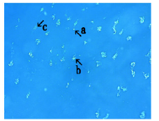
Figure 1: Microscopic appearance of synthetic DNA (E. coli) crown cells without salt. DNA synthetic crown cells were incubated for 0.5 h at 37C. The figure shows a typical single synthetic DNA crown cell (arrow a). Cells are round, typically 2 to 3 μm in diameter, and sometimes surrounded by a fuzzy layer (layer like cloud). Clusters containing two synthetic DNA crown cells are shown (arrow b). A fuzzy object without cells is shown (arrow c).
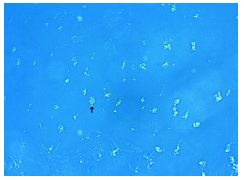
Figure 2: Microscopic appearance of synthetic DNA (E. coli) crown cells after 1.5 h at 37 C. Several fuzzy objects are shown. A fuzzy object containing several 2 to 3 μm cells is shown (arrow).
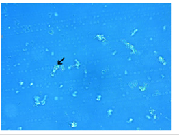
Figure 3: Microscopic appearance of synthetic DNA (E. coli) crown cells at 3.0 h. Cell descriptions as for Figure2. Many cells (2 to 3 μm) within fuzzy object were observed (arrow).
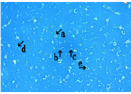
Figure 4: Microscopic appearance of synthetic DNA (E. coli) crown cells at 18 h. Objects with various shapes and sizes are shown. An object formed a complete ring (arrow a). Another object formed an incomplete ring (arrow e), which other objects formed partial rings (arrows b and c). A single synthetic DNA crown cell (2 to 3 m) is shown (arrow d).
As described previously [9,11,12], these cells were synthesized
as follows:
Step 1: Sph binds to DNA and forms thin fibers.
Step 2: The fibers formed a bunch together with A-M
compounds.
Step 3: Loops of the bunch (synthetic DNA crown cells) were
formed with monolaurin. Therefore, objects
(Figure 4, arrows b, c and d) may represent different steps in cell formation. (Figure 4, arrow a) shows a complete ring; based on these microscopic observations, this ring was a synthetic DNA crown cell. However, it is not clear whether such cells were the same as those that were prepared in previous experiments. Figure 1 to 4 show the loose assembly, or cell population-like cluster, that was spontaneously formed among synthetic DNA crown cells. It is possible that new cells (regenerated synthetic DNA crown cells) were generated from these assemblies.
Assembly formation in mixtures of synthetic DNA (E. coli) crown cells with salt A after incubation for 0.5, 3.0 and 18.0 h incubation at 37°C
When salt A was combined with DNA crown cells and observed for 0.5 h, several kinds of assemblies (varying in size and shape) were observed; one such assembly is shown in Figure 5. The assembly consists of several fuzzy objects (Figure 5, arrow a). Streak-like objects were also observed within the assembly (Figure 5, arrow b). A synthetic DNA crown cell measuring approximately 2 to 3μm is shown in (Figure 5, arrow c). Figure 6 shows the appearance of synthetic DNA (E. coli) crown cells after culturing with salt A for 3.0 h. A rock-like assembly, possibly consisting of several assemblies, is shown in (Figure 6, arrow a). Also, several ring-like objects were observed within the assembly (Figure 6, arrow b). A fuzzy object containing about 10 cells (each cell was approximately 2 to 3 μm) was observed (Figure 6, arrow c). Figure 7 shows the cultures 18 h after salt addition; the assembly consists of numerous irregular objects. The arrow in Figure 7 shows a bunch of Sph-DNA fibers that were used for cell formation. Several objects (Figure 7, arrows a, b, c and d) were observed in each bunch; these objects were used to construct the outside of the cells and rings (Figure7, arrows a and d). Rings were the same modified synthetic DNA crown cells shown in Figs. 1 to 4. Here, these ring-like objects were called modified synthetic DNA crown cells.
In summary, synthetic DNA crown cells were produced as
follows:
• Formation of a bunch of fibers (Figure 7, arrow)
• Formation of a ring-like bunch (Figure 7, arrows a, b and c)
• Formation of synthetic DNA crown cells (complete ring) by the
bunch rotating (Figure 7, arrow d).
Also, a large ring was observed (Figure 7 arrow e); however, it is studying on the relationship of the cell generation. A single DNA crown cell (approx. 2 to 4 μm) is shown in (Figure 7, arrow f).
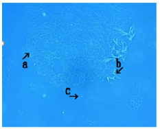
Figure 5: Microscopic appearance of DNA (E. coli) crown cells incubated in Salt A for 0.5 h at 37 C. Assembly consists of several objects with fuzzy substances (arrow a). Rod-like cells are also visible within the assembly (arrow b). A synthetic DNA crown cell (approx. 2 to 3 m) is shown (arrow c).
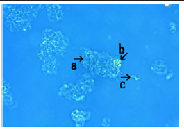
Figure 6: Microscopic appearance of synthetic DNA (E. coli) crown cells in Salt A for 3 h at 37 C. Each assembly had a rock-like appearance, with several assemblies as shown (arrow a). Several ring-like objects are visible within the assembly (arrow b). A fuzzy object containing about 10 cells (each cell was approx. 2 to 3 m) was observed (arrow c).
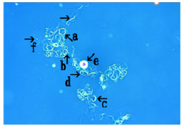
Figure 7: Microscopic appearance of synthetic DNA (E. coli) crown cells in salt A for 18 h at 37 C. Assembly consisting of many irregular objects in a bunch (arrow, arrows; a, b, c, and d). A large ring is also shown (arrow e). A single DNA crown cell is shown in Figure7 (arrow f; approx. 2 to 4 μm).
Microscopic appearance of synthetic DNA (E. coli) crown cells after incubation with salt B for 3.0 and 18.0 h
The assembly formed after incubation of synthetic DNA (E. coli) crown cells in salt B for 3.0 h is shown in Fig. 8. These assemblies may form from many objects with fuzzy like spaces (Figure8, arrow a). However, cell-like objects were not observed within the assembly.
A single synthetic DNA crown cell was observed in Figure 8, arrow b (approx. 2 to 4 μm in size).
The assembly, which was formed after culturing of synthetic DNA (E. coli) crown cells for 18 h in salt B is shown (Figure 9, arrow a). The assembly consists of numerous cell-like objects (Figure 9, arrow b) containing irregular objects (Figure 9, arrow c). The round objects (Figure9, arrow b) were modified synthetic DNA crown cells and irregular objects (Figure 9, arrow c) consisted of Sph-DNA fibrous branches.
Also, another object was observed within the assembly (Figure9, arrow e), however, now, it was not clear whether it was related with the formation of synthetic DNA crown cells. A single synthetic DNA crown cell was observed in (Figure 9, arrow d) measuring approx. 2 to 4μm in size.
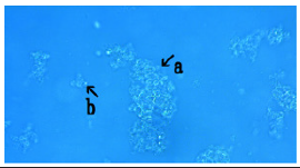
Figure 8: Microscopic appearance of synthetic DNA (E. coli) crown cells incubated in salt B for 3 h at 37 °C. The assembly formed after incubation in salt B for 3 h is shown in Figure 8. The assembly was formed of many objects with several fuzzy spaces (arrow a). A single synthetic DNA crown cell is shown (arrow b; approx. 2 to 4 m).
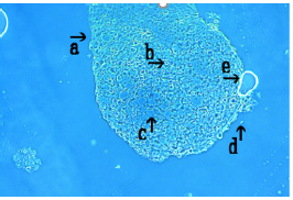
Figure 9: Microscopic appearance of synthetic DNA (E. coli) crown cells incubated in salt B for 18 h at 37 C. The assembly (arrow a) consists of many round objects (arrow b) and contains irregular objects (arrow c). A object like round was observed (arrow e). A single synthetic DNA crown cell (2 to 4 μm) was observed (arrow d).
Microscopic features of synthetic DNA crown cells incubated in salt C for 3.0, 18.0 and 72 h at 37°C
Figure 10 shows the appearance of synthetic DNA crown cells incubated in salt C for 3.0 h.
Many synthetic DNA crown cells were observed within fuzzy layer (Figure 10, arrow a). In addition to a typical cell-like round body measuring approximately 10μm in size was also observed (Figure 10, arrow b), a DNA crown cell measuring approx. 2 to 4 μm was observed (Figure 10, arrow c). Figure11 shows the appearance of synthetic DNA (E. coli) crown cells after incubation in salt C for 18.0 h; an assembly consisting of irregular objects was observed (Figure 11). Figure 12 shows the appearance of synthetic DNA (E. coli) crown cells after incubation in salt C for 72 h. The assembly consists of irregular round or rod-like structures. All these objects consist of Sph-DNA fibers or bunches of fibers. Figure12 shows a microscopic appearance, which could possibly be used to explain the development of modified synthetic DNA crown cells. Arrows a and c show a process in which a ring of synthetic DNA crown cells was formed together with a bunch of Sph-DNA fibrous. Modified synthetic DNA crown cells (complete rings) can be seen in (Figure12, arrow b). A single synthetic DNA crown cell was observed in Figure12 (arrow d; approx. 2 to 3μm).
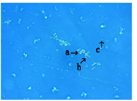
Figure 10: Microscopic appearance of synthetic DNA (E. coli) crown cells incubated in salt C for 3 h at 37 C. Many synthetic DNA crown cells can be seen in the fuzzy space (arrow a). A typical round object (approx. 10 m) is shown (arrow b). A synthetic DNA crown cell is shown (arrow c; approx. 2 to 4 m).
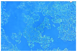
Figure 11: Microscopic appearance of synthetic DNA (E. coli) crown cells incubated in salt C for 18 h at 37 C. An assembly consisting of irregular objects is shown.
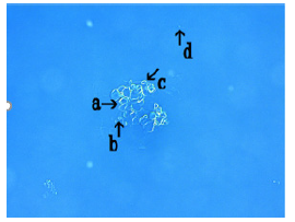
Figure 12: Microscopic appearance of synthetic DNA (E. coli) crown cells incubated in salt C for 72 h at 37 C. The assembly consists of round or rod-like objects, as well as bunches (arrows a and c). Ring-like objects are also shown (arrow b). A single synthetic DNA crown cell is shown (arrow d; approx. 2 to 3 m).
Microscopic features of synthetic DNA crown cells incubated in salt D for 3.0 and 72 h at 7°C
Figure 13 showed the appearance of synthetic DNA (E. coli) crown cells after the addition of salt D for 3.0 h.
A large assembly was observed (Figure 13, arrow a); a round object (arrow b) and a rod (arrow c) were observed within the assembly. A single synthetic DNA crown cell was observed in (Figure 13, arrow d); approx. 2 to 3μm). Figure 14 shows the appearance of synthetic DNA (E. coli) crown cells after the addition of salt D for 72 h. A cluster consisting of several synthetic DNA crown cells (round objects) was observed (Figure 14, arrow a), and a single cell was observed in (Figure 14, arrow b). Further, synthetic DNA crown cells (rod-like objects) were observed (Figure 14, arrow c). A single DNA crown cell was observed in (Figure 14, arrow d); approx. 2 to 3μm). Before the experiments using salt were performed, experiments were carried out using synthetic DNA crown cells without salt. Interestingly, the loose assemblies or clusters of synthetic DNA crown cells were formed spontaneously when such cells were cultured. After 18 h of culture, round objects (Figure 4) were observed. Experiments were then carried out using several commercial salts, which contained NaCl, CaCl2, MgCl2 and KCl without any carbohydrates, proteins and/or lipids. However, the rate of their substances was different for each salt. When salts were added to synthetic DNA crown cells and the mixtures were cultured at 37°C, the assembly was formed within 0.5 h in all salts tested and the assembly became large over time. It is unclear whether the different salts had different effects on the development of the assembly. Objects like rods were formed using salt D, which has low NaCl content.
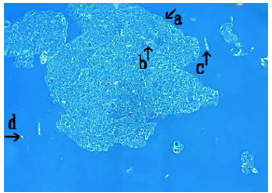
Figure 13: Microscopic appearance of synthetic DNA (E. coli) crown cells incubated in salt D for 3 h at 37 C. A large assembly is shown (arrow a). A round object was observed within the assembly (arrow b). Also, rod-like objects are shown in the assembly (arrow c). A single synthetic DNA crown cell is shown (arrow d; approx. 2 to 3 m).
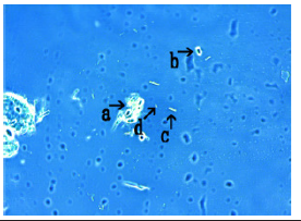
Figure 14: Microscopic appearance of synthetic DNA (E. coli) crown cells incubated in salt D for 72 h at 37 C. A cluster consisting of several round objects is shown (arrow a). Also, a single cell is shown (arrow b). Rod-like object is shown (arrow c). A single DNA crown cell is shown (arrow d; approx. 2 to 3 m).
However, generally, assemblies are considered to develop as
follows:
Step 1: Fuzzy layer with synthetic DNA crown cells developed
during incubation.
Step 2: Binding among the developed synthetic DNA crown
cells occurred spontaneously or with salt and a cluster
(population) of synthetic DNA crown cells were formed.
Step 3: Next, binding among such clusters (populations) of
synthetic DNA crown cells occurred and an assembly was
formed (referred to here as a developed assembly).
Developed assemblies may be formed based on the adsorptive characteristics of synthetic DNA crown cells. In addition, round objects were observed in most of the developed assemblies. Although partially described above, such objects were modified synthetic DNA crown cells based on the following circumstantial evidence. To date, assemblies have been reported to be formed by co-culturing synthetic DNA crown cells with Bacillus subtilis or yeast. In many cases, round objects were observed within the assembly [11,13- 15]. However, such objects could not be considered as cells, because living organisms (Bacillus or yeast) were contained in the culture. On the other hand, in present experiments, no living organisms were used; rather, the only materials that were used were Sph, DNA, adenosine and monolaurin. Therefore, all round objects were synthetic DNA crown cells, and they may be modified within the assembly and referred to as modified synthetic DNA crown cells.
The findings indicated that modified synthetic DNA crown cells were produced within the assembly.
In this paper, synthetic DNA crown cells, modified synthetic DNA crown cells, regenerated synthetic DNA crown cells and DNA crown cells were used differently. Synthetic DNA crown cells were used as experimental materials; modified synthetic DNA crown cells were round objects, which were observed within the assembly, regenerated synthetic DNA crown cells were round objects that were separated from the assembly, and DNA crown cells were cells which multiplied within egg white. These cells have basic structures of DNA crown cells, with an outer membrane that consists of DNA. However, except for these, it is unclear whether there were any differences between synthetic DNA crown cells, modified synthetic DNA crown cells, generated synthetic DNA crown cells and DNA crown cells.
In summary, the generation of new DNA crown cells occurs as follows:
First, the assembly of synthetic DNA crown cells, which includes the reconstruction of synthetic DNA crown cells, occurred. Second, synthetic DNA crown cells were modified within the assembly (referred to in this study as modified synthetic DNA crown cells). Third, modified synthetic DNA crown cells are released from the reconstructed assembly and synthetic DNA crown cells are produced (referred to in this study as regenerated synthetic DNA crown cells).
However, several problems remain regarding regeneration of
synthetic DNA crown cells:
• If modified or regenerated synthetic DNA crown cells were
the products, it is unclear whether these cells could selfreplicating.
• Most modified synthetic DNA crown cells were larger than
synthetic DNA crown cells (original synthetic DNA crown
cells), measuring at least 10 μm in size. Therefore, regenerated
synthetic DNA crown cells may be produced from modified
synthetic DNA crown cells.
• In previous experiments on assembly formation, most round
objects were released from the assembly [14,15]. In the
present experiments, most modified synthetic DNA crown
cells remained within the assembly.
To resolve these problems, I will conduct experiments using specific inorganic materials, such as NaCl and KCl. Such experiments will provide more information on the regeneration of synthetic DNA crown cells.
References
- Inooka S (2012) Preparation and cultivation of artificial cells. App Cell Biol 25: 13-18.
- Inooka S (2016) Preparation of Artificial Cells Using Eggs with Sphingosine-DNA. J Chem Eng Process Technol 7(1): 277.
- Inooka S (2016) Aggregation of sphingosine-DNA and cell construction using components from egg white. Integrative Molecular Medicine 3(6): 1-5.
- Inooka S (2017) Biotechnical and Systematic Preparation of Artificial Cells. The Global Journal of Research in Engineering 17(1).
- Inooka S (2017) Systematic Preparation of Bovine Meat DNA Crown Cells App. Cell Biol Japan 30: 13-16.
- Inooka S (2018) Systematic Preparation of DNA (Akoya pearl oyster) Crown Cells App. Cell Biol Japan 31: 21-34.
- Inooka S (2019) Preparation of DNA (Nannochloropsis species) Crown Cells Using eggs, sphingosine and DNA, and subsequent cell recovery App. Cell Biol Japan 32: 55-64.
- Inooka S (2014) Preparation of Artificial Human Placental Cells App. Cell Biol 27: 43-49.
- Inooka S (2017) Systematic Preparation of Artificial Cells (DNA Crown Cells) J. Chem Eng Process Technol 8(2): 327.
- Inooka S (2021) The assembly formation by co-cultures of DNA (Escherichia coli) crown cells with Bacillus subtilis cells. Journal of Biotechnology & Bioresearch 2(5).
- Inooka S (2021) Microscopic observation of assemblies formed in mixtures of DNA (human placenta) crown cells with Bacillus subtilis. An archive of organic and in-organic chemical sciences. 5(2): 658-664.
- Inooka S (2018) Theory of cyto-organisms generation –Artificial cells produced with egg; the discover and synthesis of DNA crown cells. (In Japanese) Daigaku Kyouko Press Okayama Japan.
- Inooka S (2021) Synthetic DNA Crown Cells kill Yeast: Relationship with Assembly Formation of Yeast. Civil Engineering Research Journal 12(2).
- Inooka S (2021) Assembly and Bacterial Growth Suppression in the Co-culture of Synthetic DNA (Akoya Pearl Oyster) Crown Cells with Bacillus subtilis. Applied Cell Biology Japan 4: 67-84.
- Inooka S (2022) Assemblies in Synthetic DNA (Nannochloropsis Species) Crown Cells with Bacillus subtilis. Acta Scientific Microbiology 5(1): 03-08.


 We use cookies to ensure you get the best experience on our website.